|
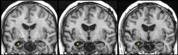
| Figure 2: Reduction of lateral volume in the HC head and/or body in this TBI patient is clearly indicated when the IHLV (yellow asterix) has enlarged or appears large. Left=acute, Middle=6 months post-TBI, Right=12 months post- TBI. Although this represents an extreme case of atrophy for the illustration of IHLV enlargement, this was necessary to illustrate the relationship between IHLV enlargement after TBI, and time. |