 |
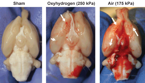
| Figure 1: Qualitative examination of vascular damage can be observed in the images above depicting hematoma and petechial hemorrhage on the ventral surface of brains taken at 3 hours post-injury from rats exposed to oxyhydrogen- or compressed air-driven shockwaves. Note larger hematoma and more extensive petechial hemorrhage (white arrows) in the brain from a compressed air-driven shockwave-exposed rat. Images are representative of six rats per treatment group. |