 |
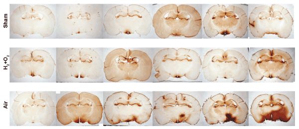
| Figure 2: Histological evidence of blood-brain barrier compromise following compressed airdriven shockwave exposure. IgG extravasation can be visualized in the inferior left (side of the shockwave source) side of the brains in 5 of 6 rats exposed to compressed air-driven shockwaves. This region specific staining is not evident in the brains of rats exposed to sham injury or oxyhydrogen-driven shockwaves. Brain sections are each from a different animal euthanized at 3 hours post-injury and are presented in order from least severe to most severe IgG staining. n=6 per treatment. |