|
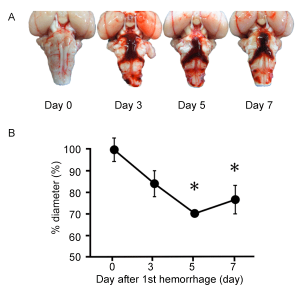
| Figure 1: Clot formation and time course of narrowing of the rabbit basilar artery after SAH. (A) Representative photographs of the ventral surface of the rabbit brain removed on days 0, 3, 5, and 7 after subarachnoid hemorrhage (SAH). (B) Time course of changes in the external diameter of rabbit basilar arteries after SAH. Data are the means ± s.e.m. (n = 3, each time point). The external diameter on day 0 was assigned as 100%. * P<0.05 versus day 0. |