|
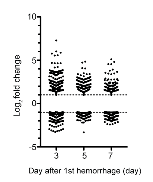
| Figure 3: Scatter diagram of differentially expressed genes in the rabbit basilar artery after SAH. Plots in the scatter diagram showed individual up- and down-regulated genes on days 3, 5 and 7 compared to day 0. The broken lines indicated the threshold of log2 fold change. The upper broken line indicates log2 fold change = 1, and the lower one indicates log2 fold change = −1. |