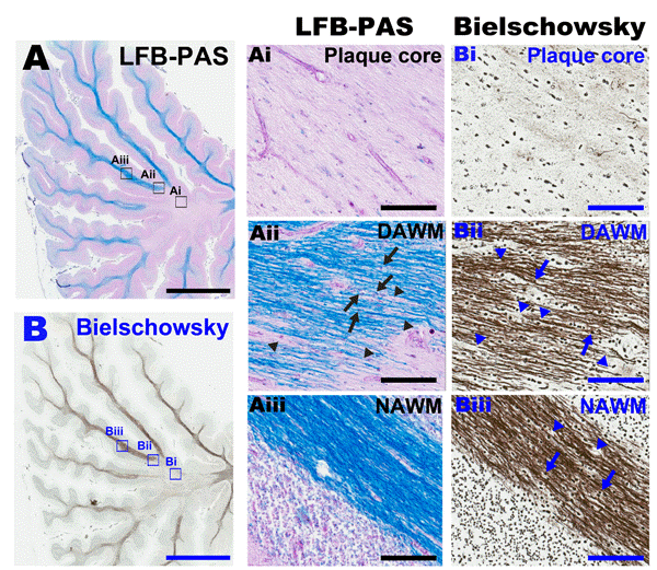
Figure 1: Chronic-active MS plaque in the cerebellar white matter
and mechanisms of MS pathology (A) Luxol fast blue with periodic
acid-Schiff's (LFB-PAS) histochemistry stain of cerebellar white
matter in a secondary-progressive MS patient. Higher
magnification shows the different pathological regions involving
neurodegeneration (Ai-Aiii); (Ai) plaque core shows complete loss
of myelin; (Aii) Dirty-appearing white matter (DAWM) shows
demyelination with LFB pallor (arrowheads) with differential
staining of infiltrating macrophages (arrows); (Aiii) Normalappearing
white matter (NAWM) shows intact myelin integrity. (B)
Bielschowsky histochemistry of a serial section of cerebellum from
the same patient. (Bi) Plaque core shows complete loss of axons
demonstrated by lack of silver staining; (Bii) DAWM shows axonal
damage (arrowheads) with infiltrating macrophages (arrow), (Biii)
NAWM shows less axonal damage compared to DAWM
(arrowheads) with macrophages (arrow); scale bar for low
magnification = 5mm; scale bar for high magnification = 50 m m |

