 |
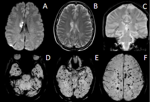
| Figure 3: The present MRI of the brain 7 years after whole brain irradiation. (A) Diffusion weighted image (DWI) demonstrates restricted diffusion at the head and body of right caudate nucleus and globuspallidus. (B) T2 weighted image (T2WI) demonstrates spots of low signal intensity areas in the subcortical and deep white matter of both hemispheres. (C) GRE T2*WI demonstrates blooming dark signal spots in the subcortical and deep white matter areas of bilateral hemispheres. (D,E,F) Susceptibility weighted image (SWI) shows numerous spots of micro hemorrhage in subcortical and deep white matters of the bilateral cerebral hemisphere. |