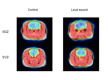
Figure 3:
An example of coronal PET images superimposed on MRIs of the SGZ (upper) and SVZ (lower) regions of the rat brains (left: control, right: loud noise). The dashed lines indicate regions of interest.