 |
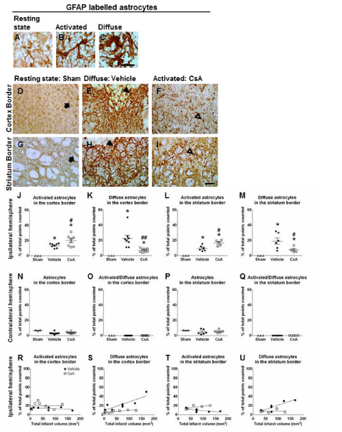
| Figure 2: Effects of CsA treatment on activated versus diffuse astrocytes and correlation with infarct volume. Bright-field immunohistochemical photomicrographs depicting GFAP staining of resting astrocytes within the ipsilateral hemisphere of shamoperated controls (A, D), diffuse astrocytes (B, E) and activated astrocytes (C, F) within the border cortex and striatum of the ipsilateral hemisphere from vehicle and CsA treated animals using consistent sample areas. Short arrows represent cells in resting states with long slender processes and low level of GFAP expression, open arrow heads indicate activated astrocytes with cellular hypertrophy up-regulation of GFAP without process overlap, and filled arrow heads represent diffuse astrocytes with cellular hypertrophy and pronounced process overlap. Scatter plots demonstrate CsA significantly increased the number of activated and decreased the number of diffuse astrocytes within the border cortex and striatum (G-J, ANOVA). Data are presented as mean ± SEM, *P<0.05 compared to sham-operated animals, #P<0.05, ##P<0.005 compared to vehicle treated animals. No difference in the number of resting, activated/diffuse astrocytes in the contralateral hemisphere across all groups analysed (L-N) (ANOVA). No correlation between infarct volume and the number of activated astrocytes in either treatment groups within the cortex border (O). A significant correlation was found between infarct volume and the number of severely diffuse astrocytes within the border cortex for both treatment groups (vehicle: r=0.85, P<0.01, n=8, CsA: r=0.81, P<0.05, n=8; P). No correlation was observed between infarct volume and the number of activated astrocytes in either treatment groups within the striatum border (Q). A positive correlation was found between volume of infarct and diffuse astrocyte numbers within the border striatum of vehicle treated animals only (vehicle: r=0.86, P<0.05, n=5; R) (Pearson product moment correlation coefficients). Scale bars A-F = 100μm. CsA: Cyclosporine A; GFAP: glial fibrillary acidic protein. |