|
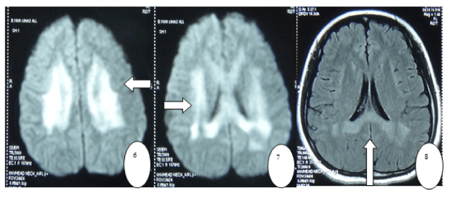
| Figures 6-8: In April 11th, 2012 brain MRI showed hyperintense T1 and T2 signals of bilateral centrum semiovale (6), callosum and white matter around rear corner of di-lateral ventricle (7), as well as hyperintense signal in diffusion-weighted imaging (DWI)(8). |