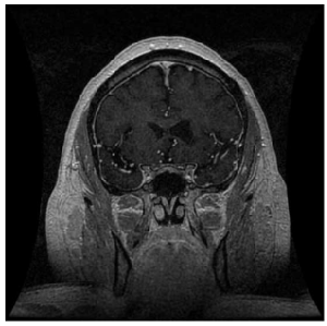
Figure 2:
Post-contrast T1-weighted MRI image demonstrating leftventricular dilatation due to atrophy, suggestive of past basal ganglia stroke.