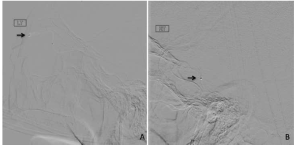
Figure 3:
Intraprocedural DSA images of the micro catheter tip position in A. Left superior ophthalmic vein and B. right cavernous sinus.