|
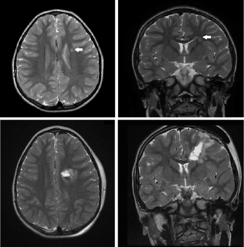
| Figure 1: Pre-operative MRI brain T2 axial and coronal images with arrows designating left frontal cortex heterotopic grey matter within the sub ependymal white matter of the left lateral ventricle and corona radiata and immediate post-operative MRI brain T2 axial and coronal images showing left frontal craniotomy with resection of heterotopia. |