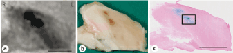
(a) Two CMBs of the right thalamus Postmortem GRE-MRI. (b) Macroscopic appearance (brown spots). (c) Iron stain (blue lesions). These CMBs are roughly similar in size in each picture. Scale bar=10 mm.
|
| Figure 2: Histopathology-neuroimaging correlation of CMBs. (a) Two CMBs of the right thalamus Postmortem GRE-MRI. (b) Macroscopic appearance (brown spots). (c) Iron stain (blue lesions). These CMBs are roughly similar in size in each picture. Scale bar=10 mm. |