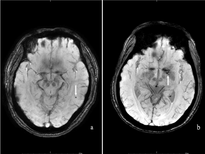
(a) Axial SWAN images depicting CMBs at corticomedullary in an mTBI patient. (b) In contrasts, CMBs associated with hypertensive vascular disease are often located at deep gray matter.
 |
| Figure 3: SWMRI imaging of CMBs. (a) Axial SWAN images depicting CMBs at corticomedullary in an mTBI patient. (b) In contrasts, CMBs associated with hypertensive vascular disease are often located at deep gray matter. |