 |
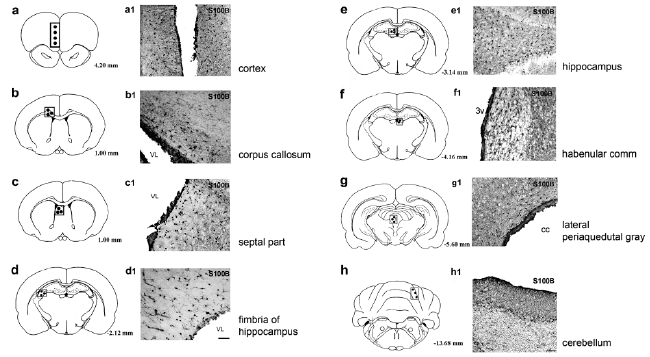
| Figure 2: Bright-field photomicrographs showing expression of the S100B protein (indirect immunohistochemistry) along the rostrocaudal axis of the adult male rat brain. S100B-IR in different brain areas revealed a heterogeneous pattern of cell population with the strongest density in the periventricular areas: (b1): the corpus callosum, (c1): lateral part of the septal area, (d1): the fimbria of the hippocampus, (f1): the habenular comn, (g1): lateral periaquedutal gray. The cortical area adjacent to longitudinal fissure (a1), the central region of hippocampus between dentate gyrus (e1) and specific cerebellar layers (h1) also showed a high density of cells that were S100B-IR. Schematic drawings in a, b, c, d, e, f, g and h were obtained from Paxinos and Watson, 1998. The squares indicate the respective magnified areas. Scale bar: 100 μm. |