|
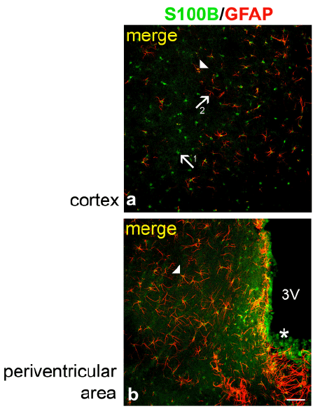
| Figure 4: S100B IR and GFAP-IR in the cortex and periventricular areas of Wistar rats. Arrow 1 indicates S100B-IR cells (green), Arrow 2 indicates GFAP-IR cells (red) a, and the arrowheads indicate double-immunoreactive cells (yellow) observed in the cortex (a) and periventricular areas (b). Note that in contrast to the periventricular areas, the colocalization of S100B-IR in GFAP-IR cells was lower in cortical areas (a). In b asterisk indicates S100B-IR ependymal cells. Scale bar: 100 μm. |