|
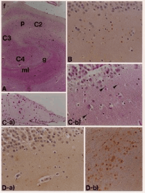
| Figure: 1 (A): The hippocampal formation stained by Hematoxilin Eosin. g: granular layer p: pyramydal neuron ml: molecular layer f: fimbria (B): SPDs detected by lectin stains. GSI-B4. (C-a): Corpora amylacea are intensely stained by PAS. (C.b): SPDs are weakly stained by PAS. (D-a): SPDs show intense reactivity with antichondroitin sulfate. (D-b): Corpora amylacea show intense reactivity with anti-tau protein antibody. |