|
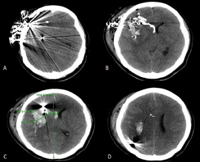
| Figure 1: Axial CT brain showing the bullet entry wound in the right frontotemporal region; the bullet did not exit the intracranial cavity (A). Several bullet fragments are lodged in the tissue of the right frontal lobe. At the level of the lateral ventricles there is evidence of bony fragments in the overlying scalp tissue and right frontal lobe extending into the right lateral ventricle (B). A large intraparenchymal hematoma is present in the right frontal lobe adjacent to additional bullet and bone fragments with approximately 9mm of midline shift (C). Additional bullet fragments are appreciated in the right parietal lobe with a small subdural hematoma overlying the right cerebral hemisphere (D). Diffuse sulcal effacement is appreciated in all images. |