|
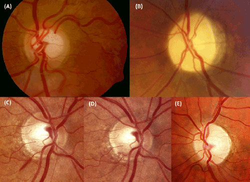
| Figure 2: Differences between glaucomatous optic neuropathy and NGON. A. Glaucomatous optic neuropathy: Neuroretinal ring has a good healthy color but appears markedly thin, especially in superior. Enlargement of the cup is obvious. Parapapillary atrophy is shown in the nasal and temporal borders. This is a classically sign of glaucomatous optic disc, but it can also be seen in elderly patients. We also can see the vessels displaced to the nasal side and a notch in the inferior border. B. NGON: Neuroretinal ring has a pronounced pallor, showing a whole atrophy of the optic disc. This picture corresponds to a past NAION. C, D. Evolution of a disc hemorrhage in the inferior border in a glaucomatous disc. Usually disappears in 2- 6 months, and means progression of the optic nerve head damage. E. Optic atrophy in a NGON with superior and temporal pallor of neuroretinal ring. Courtesy of Spanish Society of Ophthalmology. |