|
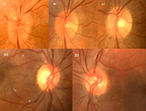
| Figure 3: Superior: Evolution of an anterior MS- ON. A. Optic nerve head edema in the acute phase with reduction of VA. B. Reduction of edema. C. Complete resolution of the edema with VA reconstitution. Inferior: D. Pallor of the optic disc in the right eye with past anterior MS- ON. It is especially manifest in temporal side of neuroretinal ring when we compare it with the fellow eye (E). Courtesy of Spanish Society of Ophthalmology. |