|
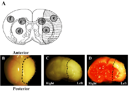
| Figure 1: Rat model of neonatal hypoxic-ischemic encephalopathy (HIE) (A) The dotted area represents the infarct region. Areas a and c, but not b, are infarct regions. (B-D) Stereomicroscopic images of the brain of a Wistar rat 3 days after HIE induction. Dashed lines indicate the infarct area. (B) Whole brain; (C) coronal section; (D) triphenyltetrazolium chloride (TTC)-stained coronal section; infarct area is not stained. |