|
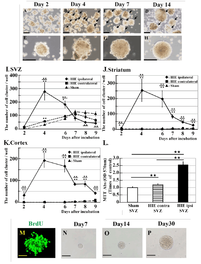
| Figure 2: Formation of neurosphere-like cell clusters by cells harvested from HIE model animals. (A-H) Cells obtained from the infarct cortex form cell clusters in neurosphere culture assays that increase in diameter over time. Images were obtained after 2, 4, 7, and 14 days in culture. Scale bars: 200 μm (A-D) and 100 μm (E-H). (I-K) Numbers of cell clusters greater than 50 μm in diameter in neurosphere cultures derived from areas a-f (see Figure 1) on culture day 9. (I) Subventricular zone (SVZ); (J) striatum; (K) cortex. HIE ipsilateral and contralateral areas: n=6; sham-operated: n=6. Statistical comparisons among groups (1. sham-operated 2. HIE contralateral side 3. HIE ipsilateral side) were performed using the Games-Howell test or Mann-Whitney U test. Data are shown as mean ± SEM. ★★:1 vs. 3, ★★ or ★:1 vs. 2 or 2 vs. 3. (L) Proliferation of cells in the SVZ of sham-operated animals and the contralateral (HIE contra) or ipsilateral (HIE ipsi) SVZ of HIE model animals, as measured by the MTT assay. (I-L) ★ P<0.05 and ★★ P<0.01. (M-P) Proliferating cell in the D4 cell clusters incorporated BrdU and formed secondary neurosphere-like cell clusters. (M) Proliferating cells stained for BrdU (L: green) observed in the D4 cell cluster. (N-P) Secondary neurosphere-like cell cluster formed from a single cell of the D4 cell clusters after 7, 14, and 30 days in culture. Scale bars: 100 μm. |