|
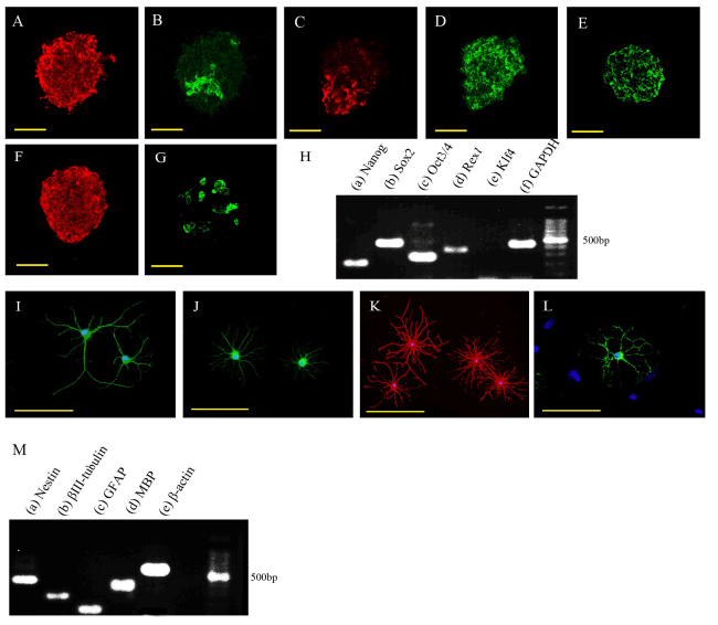
| Figure 3: Characterization of D4 cell clusters derived from the infract cortex area and differentiation of D4 cell clusters. (A-G) Immunocytochemistry of D4 cell clusters. (A) nestin; (B) neural/glial antigen 2 (NG2); (C) glial fibrillary acidic protein (GFAP); (D) β-III-tubulin; (E) oligodendrocyte marker O4; (F) vimentin; (G) ionizing calcium-binding adaptor molecule 1 (Iba1). Scale bars: 50 μm. (H) RT-PCR analyses showing expression of (a) NANOG, (b) (sex determining region Y)-box 2 (Sox2), (c) octamer-binding transcription factor 3/4 (Oct3/4), and (d) Rex1 in D4 cell clusters. Expression of KLF4 (e) was not observed. (f) Glyceraldehyde-3-phosphate dehydrogenase (GAPDH) internal control. (I-L) Immunocytochemistry of differentiated cells from D4 cell clusters. (I) β-III-tubulin (green); (J) microtubule-associated protein 2 (MAP2; green); (K) GFAP (red); and (L) O4 (green). Nuclei were counterstained with 4',6-diamidino-2-phenylindole (DAPI; blue). Scale bars: 50 μm (I, J, and L) and 100 μm (K). (M) RT-PCR analyses showing expression of (a) nestin, (b) β-III-tubulin, (c) GFAP, and (d) myelin basic protein (MBP) in differentiated D4 cell clusters. (e) β-actin internal control. |