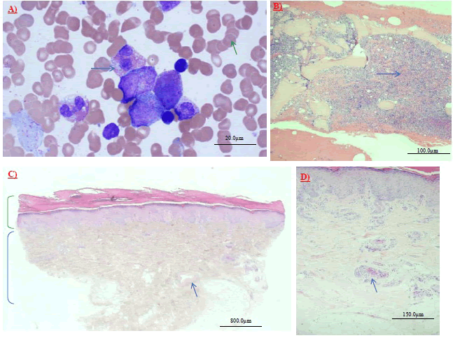
| Figure 2: (A) Bone marrow aspirate smear (high power): noted redblood cells with occasional rouleaux formation (green arrow); aswell as atypical eosinophil with clumping of eosinophils (bluearrow). (B) Bone marrow biopsy (low power); with notablehypercellular appearance; and noted mature trilineagehematopoiesis (megakaryocytes, erythroid precursors and myeloidprecursors), along marked eosinophilia (blue arrow). (C) Skinbiopsy (low power) highlighting a vessel near the right side of thisphoto (arrow). Noted the epidermis (green bracket) and underlyingdermis layers (blue bracket). The vessel demonstrate evidence offibrin thrombus formation (better visualized under high power)(blue arrow). (D) Skin biopsy (high power) demonstrating presenceof bland thrombi (fibrin thrombus) formation in the vessel ofinterest (blue arrow) |
 .
. .
.