|
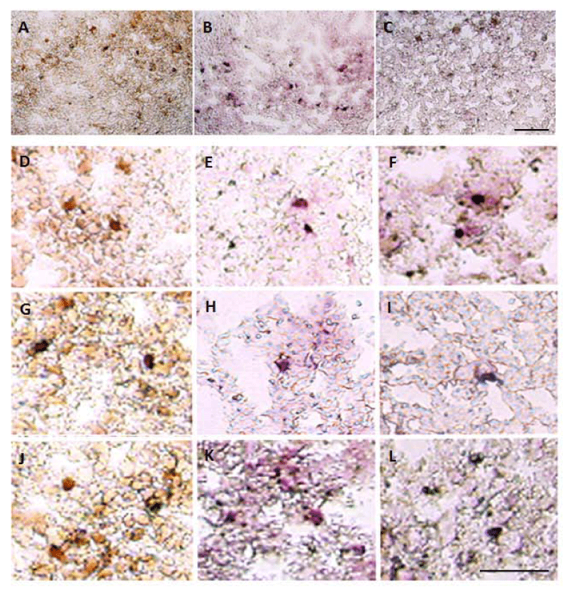
| Figure 6: Immunohistochemistry of phosphorylated tau protein and calmodulin-like skin protein (CLSP). Phosphorylated tau staining (brown color; pales A, D, G and J), CLSP staining (purple color; panels B, E, H and K) and the double staining (merged color; panels C, F, I and L). Scale bar: 50 μm. |