|
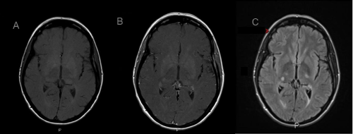
| Figure 2: (A) MRI axial brain T1 normal. (B) MRI axial brain T1 post contrast, shows a 7 mm contrast enhancing lesion within the posterior right lateral thalamus. (C) MRI T2 axial brain fluid-attenuated inversion recovery (FLAIR) shows a 7 mm hyperintensity within the posterior right lateral thalamus. |