|
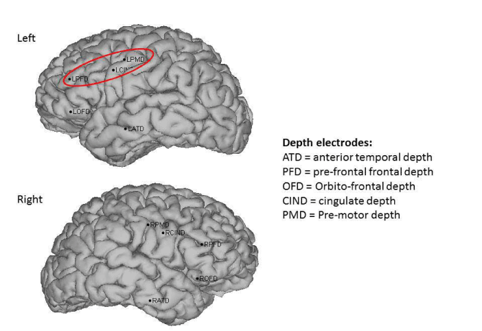
| Figure 2: Sagittal view of the positioning of the depth electrodes used for the phase II investigation projected to the patients own MRI. Five orthogonal depth electrodes were placed on each side in the orbitofrontal, prefrontal, premotor, cingulate and anterior temporal regions. The area of maximal cortical excitability identified by this investigation is circumscribed with red. |