 |
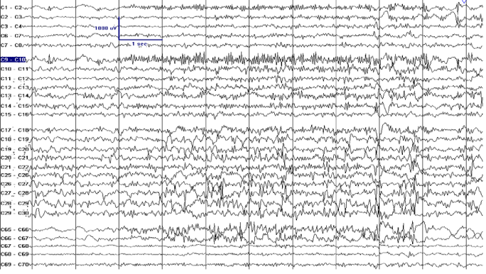
| Figure 7: Pre-resection intraoperative electrocorticography showing frequent brief bursts of polyspikes better localized at contact 1, 2, 9 and 10 that organized in runs up to 15 seconds (highlighted contacts). The electrographic signature of that region was consistent with the provisional diagnosis of focal cortical dysplasia and was confirmatory of the epileptogenic focus. |