 |
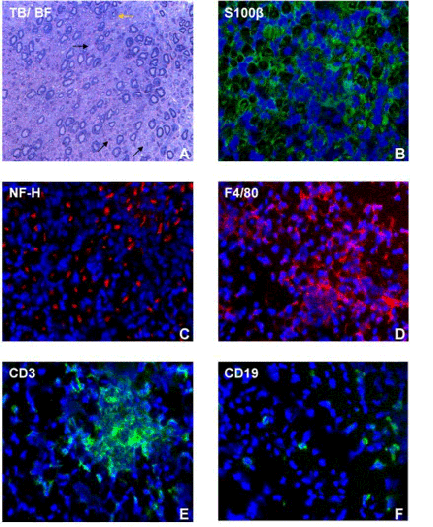
| Figure 4: Histopathology of severe murine EAN. A photomicrograph of a representative toluidine-blue stained, basic fuchsin counterstained (TB/ BF) semi-thin plastic embedded axial section of a mouse sciatic nerve shows multifocal demyelination of axons (black arrows) associated with mononuclear cell infiltrates, with reduced density of large myelinated axons (A). An endoneurial microvessel is shown (yellow arrow). Indirect immunohistochemistry photomicrographs show loss of S100ß immunoreactivity (green) associated with intense mononuclear cell infiltrate (blue), indicative of demyelination (B). There is loss of axonal density (red immunoreactivity for neurofilament-Heavy chain [NFH]) similarly associated with mononuclear cell infiltrate (blue), indicative of axonal loss in BPNM-induced murine EAN (C). Infiltrates are predominantly macrophages (F4/80+; red immunoreactivity) as shown in D. A focal collection of CD3+ T-lymphocytes and scattered CD19+ B-lymphocytes (green immunoreactivity) are shown in E and F respectively. Original magnification 400X. |