 |
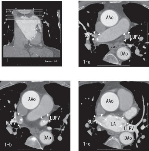
| Figure 1: Axial images showing thrombi within the right upper and the left upper pulmonary veins (white arrow head). The right upper and the left upper pulmonary veins defected most of enhancement because of full sized thrombi (1-a). The left upper pulmonary vein demonstrated the sharp merge of enhancement (1-b). The merge of the thrombi in left atrium was vague (1-c). AAo: Ascending Aorta; DAo: Descending Aorta; LA: Left Atrium; LUPV: Left Upper Pulmonary Vein; LLPV: Left Lower Pulmonary Vein; RUPV: Right Upper Pulmonary Vein. |