|
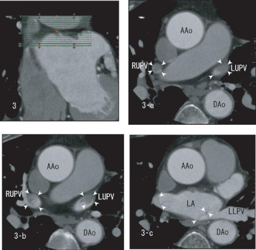
| Figure 3: After three months warfarin therapy, axial images showing the thrombi within the right upper and the left upper pulmonary veins (white arrow head). The right upper and the left upper pulmonary veins defected the most of enhancement because of full sized thrombi (3-a). The sharp borderline of the left upper pulmonary vein was disappeared and the borderline became rather vague (3-b). The size of thrombi in LA seemed to be decreased and they looked like “lines” (3-c). AAo: Ascending Aorta; DAo: Descending Aorta; LA: Left Atrium; LUPV: Left Upper Pulmonary Vein; LLPV: Left Lower Pulmonary Vein; RUPV: Right Upper Pulmonary Vein. |