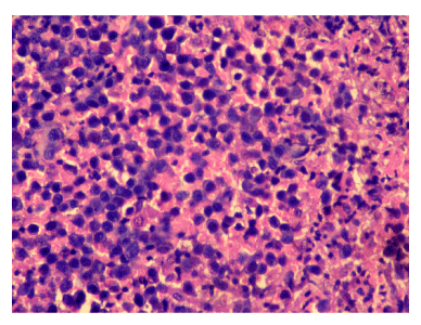
Figure 5:
Photomicrograph of histopathological analysis of biopsy specimen from conjunctival lesion showing predominantly monomorphic atypical lymphoid cells with inconspicuous nucleoli. H&E stain, 40X magnification.