|
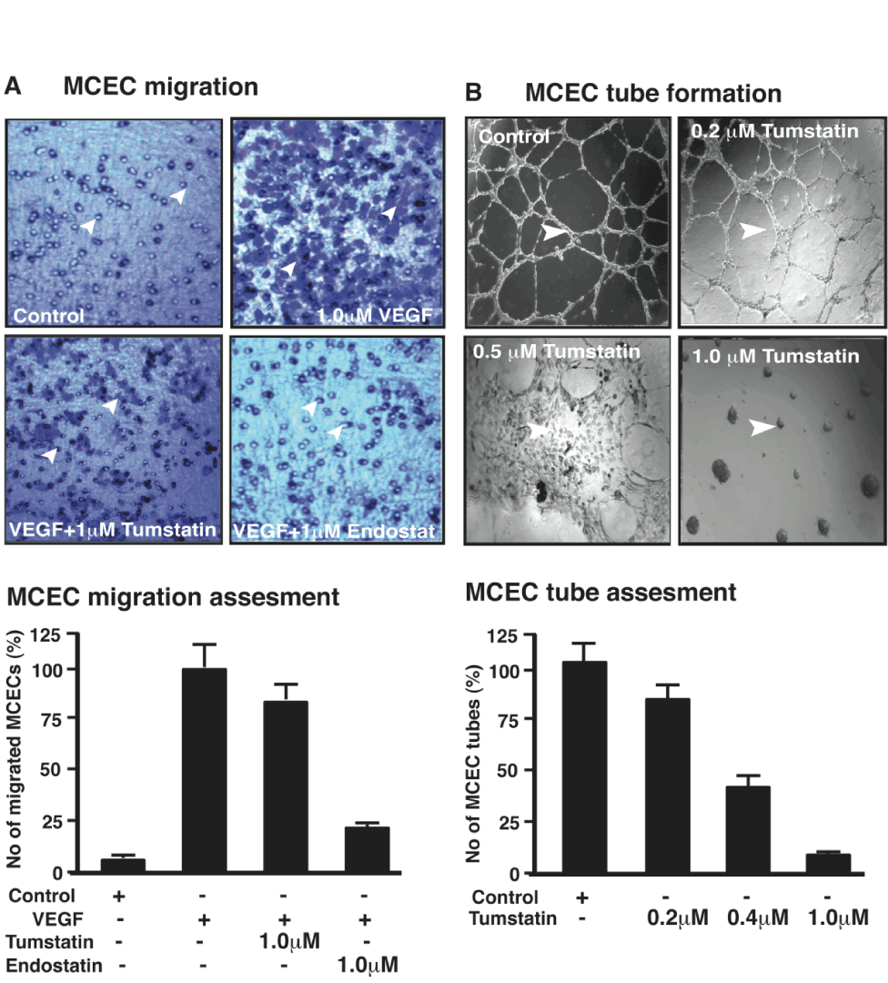
| Figure 2: Regulation of mouse corneal endothelial cell migration and tube formation by tumstatin. (A) MCEC migration. Photographs of cells from the underside of Boyden chamber membrane. Number of cells migrated in SFM (serum free medium) used as negative control. Numbers of migrated cells with VEGF or with VEGF and tumstatin or endostatin (control) as viewed using a light microscope (100x magnifications). Lower graph displays % number of cells migrated in three independent experiments. (B) Tube formation assay. MCECs treated with and without tumstatin or with endostatin (1.0µM) and seeded on Matrigel matrix. Tube formation was evaluated after 24 hr using Olympus CK2 light microscope with 100x magnification. Lower graph displays % number of tubes formed in three independent experiments. In A and B, arrows indicate migrated cells and tube formation respectively. |