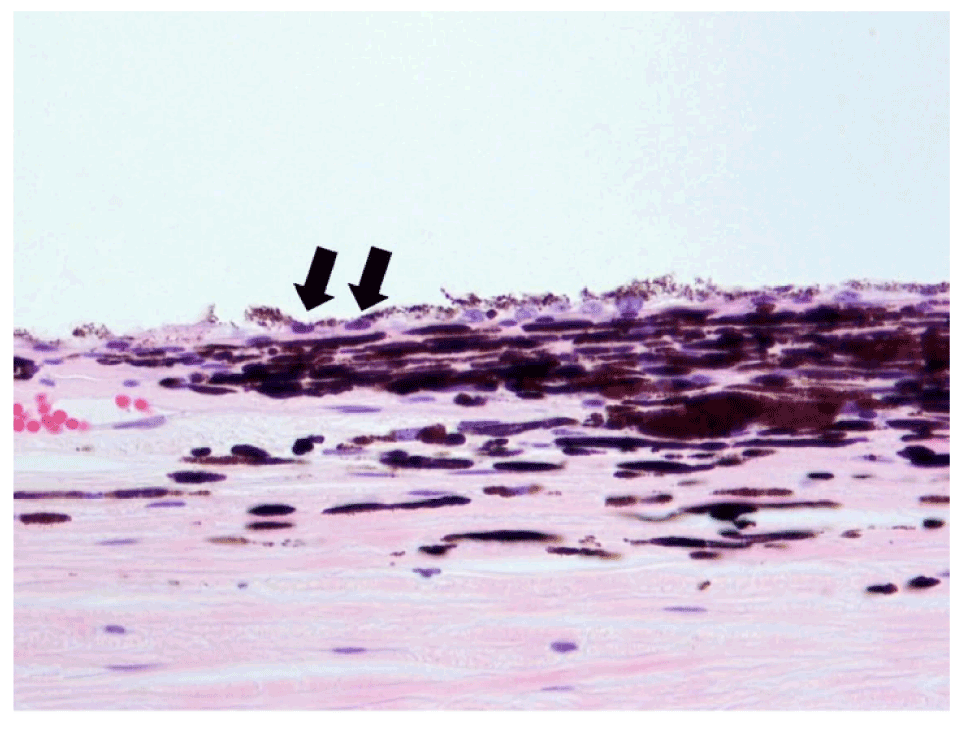
Figure 5D:
Histology of single beam 60 Gy specimen demonstrating flattened RPE cells (H&E stain).