|
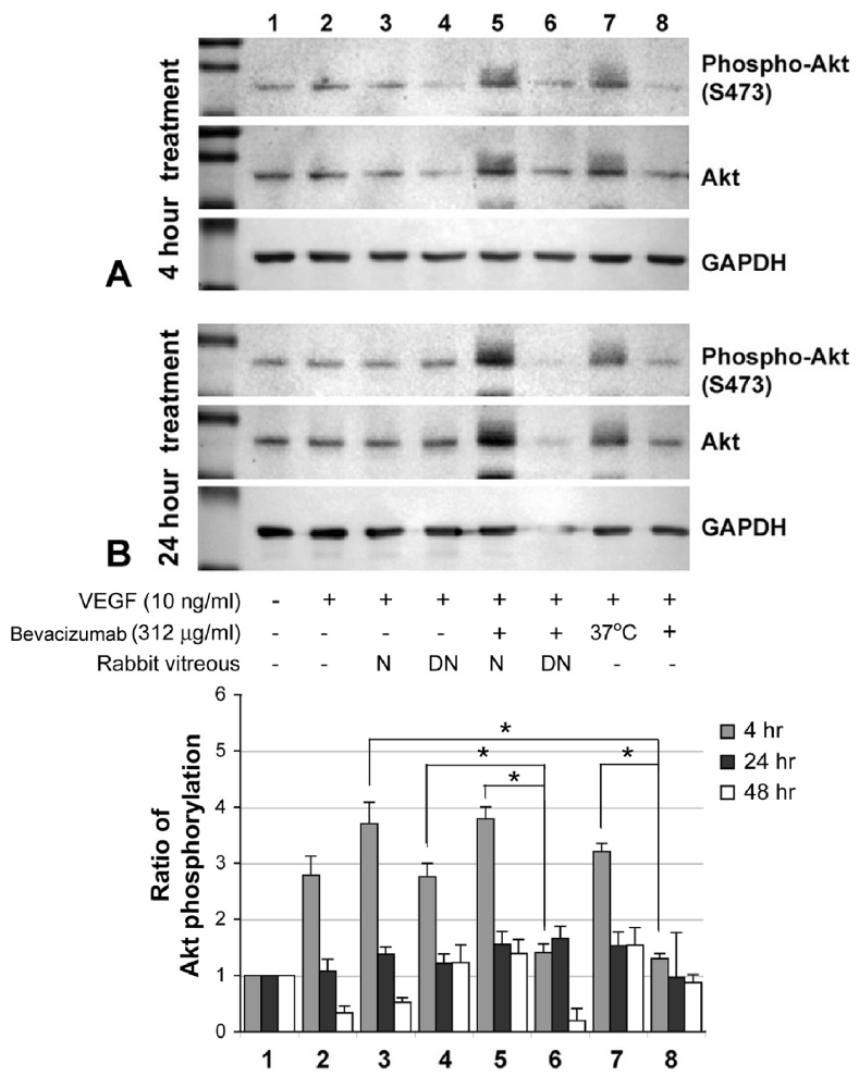
| Figure 3: Western blotting analysis of Akt activation in RF/6A cells after various treatments. (A) 4-hour treatment; (B) 24-hour treatment and (C) histogram showing the ratio of Akt phosphorylation normalized with GAPDH. Transient up-regulation of phosphorylated Akt in total Akt was resulted from VEGF (10 ng/ml) treatment. Addition of bevacizumab (312 µg/ml) blocked the Akt activation by VEGF but this effect was reduced when bevacizumab was pre-incubated with rabbit vitreous prior to addition to cells. Bevacizumab activity to block Akt phosphorylation was maintained in denatured vitreous. *indicates P<0.05, paired Students t-test. VEGF, vascular endothelial growth factor; N, native vitreous; DN, denatured vitreous; GAPDH, glyceraldehyde 3-phosphate dehydrogenase. |