|
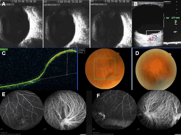
| Figure 2: Ultrasonography, OCT, Angiography, and CDFI in case 2. A Ultrasound examination showed mild vitreous opacities and posterior vitreous detachment. A dome shaped lesion of 1mm × 3mm on the peripheral globe wall with high reflective smooth surface and low internal reflectivity could be found on the 5 oclock meridian. From left to the right, the pictures showed the decreasing lesion height while a pressure was given to the globe. B By CDFI, vein flow signal with a velocity of 2.77cm/s was found inside the lesion.C Spectral domain OCT showed a crescent elevated area in the inferior fundus, large diversion of the choroidal veins could be seen converging toward this lesion. The intact neuralretinal layer and the retinal pigmental epithelial layer bulged into the vitreous in good continuity with a 1215 µm height dome shape. The reflectivity under the RPE layer was lowered. D When firm pressure was applied for seconds on the globe, the lesion collapsed. E During venous phase of ICGA, density diversions of filled choroidal vein were seen converging into a rope knot like enlarged vortex vein ampulla in the lesion. And in the corresponding FFA, slight uplift and fluorescence filling of the vortex vein ampulla could be seen. F At the area of coagulation, fluorescence dye and shelter could be found by FFA and density diversions of filled choroidal vein were also found by ICGA, but without bulging. |