|
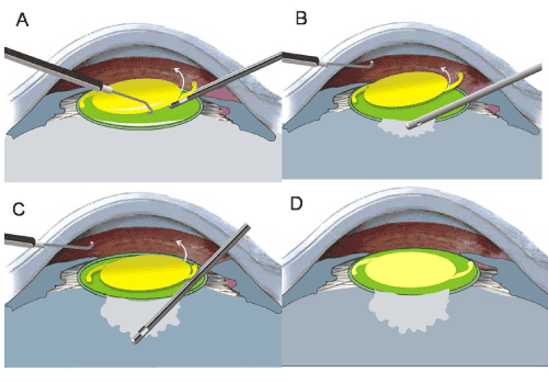
| Figure 1: 3D graphic representation of LARV showing A) a 21G curved canula causing slight decentration of the IOL to allow vitrector to pass under the optic, B) capsulovitrectomy in progress, C) anterior vitrectomy in progress, D) completed LARV with IOL secured in the bag. |