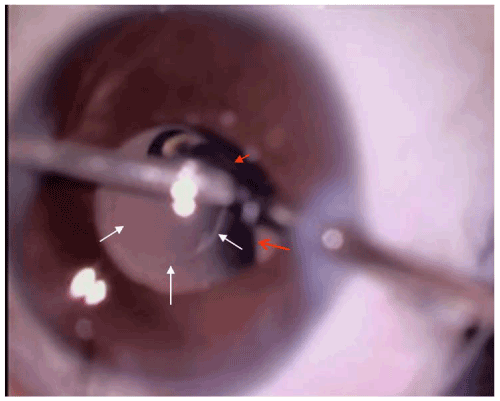
Figure 2:
Microscopic view of a LARV in progress. White arrows show margins of the posterior capsulectomy and red arrows show the margin of the IOL optic over a 21G vitrector.