|
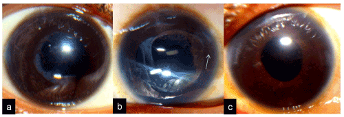
| Figure 4: Diffuse illumination digital photograph of the anterior segment showing a) IOL in the posterior chamber with iris chaffing and pigmentation on the optic, b) IOL haptic in the anterior chamber (white arrow) and c) IOL repositioned in sulcus with the optic captured behind the CCC. |