|
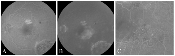
| Figure 2: (A) FA in February 2009 demonstrates telangiectasis and aneurysms in the temporal macula during the early phase. (B) Late leakage is seen on the FA done in February 2009. Magnified view of telangiectasia and aneurysms seen in the macula of panel B. |