|
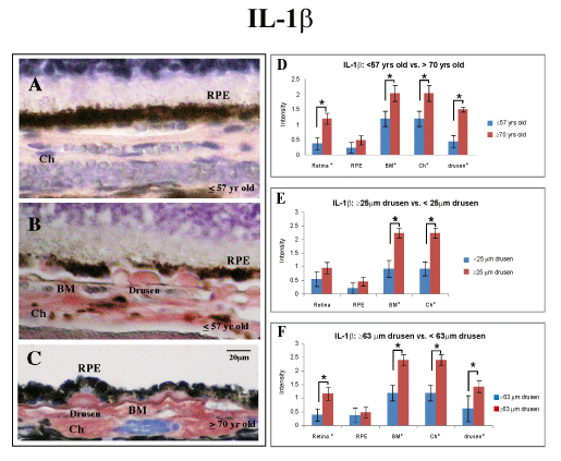
| Figure 1: Immunostaining for IL-1β with AEC development system (red) and counterstained with Mayer’s hematoxylin (blue). IL-1β expression levels in human eye tissue, analyzed by age groups and size of drusen. (A) Younger group (≤ 57 yr) with drusen ≤ 25 μm; note faint immunolabeling pattern (semi-quantitative grading of 1). (B) Eye tissue from the younger group (≤ 57 yr) with drusen ≥ 25 μm (arrows). Note immunoreactivity for IL-1β in drusen and through choroid (semi-quantitative grading of 3).RPE cells are mildly immunostained. (C) Strong immunoreactivity in drusen, Bruch’s membrane, and choroid in eyes from older group (≥ 70 yr, semi-quantitative grading of 3) with drusen ≥ 63 μm (arrows). Scale bar 20 μm. (D) Aside from the retinal pigmented epithelial (RPE) cells, tissue from older group (n = 12) are more immunoreactive for IL-1β (retinal layers through to the choroid) than younger group (n = 15) (black asterisk, p ≤ 0.05). (E) Eyes with clinically significant drusen (≥ 25 μm, n = 15) were more immunoreactive for IL-1β in Bruch’s membrane and choroid compared to eyes without clinically significant drusen (n = 12). (F) Eyes with drusen ≥ 63 μm (n = 12) were more immunoreactive for IL-1β in the retinal layers, Bruch’s membrane, choroid, and drusen than eyes with drusen ≤ 63 μm (n = 8). |