 |
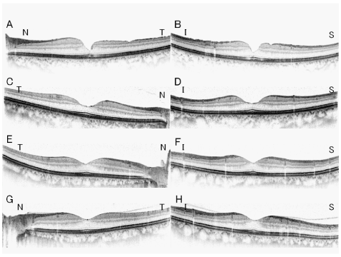
| Figure 1: Postoperative Spectralis-OCT images of a 58-year-old Japanese woman with a stage 3 macular hole (A,B) and healthy fellow eye (C, D), and those of a 48-year-old Japanese man with macula-on rhegmatogenous retinal detachment (RRD; E, F) and healthy fellow eye (G, H). A. Horizontal OCT image of a Japanese woman with a stage 3 macular hole showing a thinning of the temporal retina. Retinal ganglion cell layer (RGCL) and inner plexiform layer (IPL) are thinner which probably contributed to the thinness of the total retinal thickness in the temporal retina. B. Vertical OCT image of the same woman showing symmetrical but steep foveal contour. C. and D. In contrast, the foveal contour has a smooth concave shape in the fellow eyes. E.- H. The postoperative OCT images in eyes with RRD and fellow eyes have normal foveal contours. N, nasal; T, temporal; S, superior; I, inferior. |