 AC: Anterior Chamber
|
AC: Anterior Chamber |
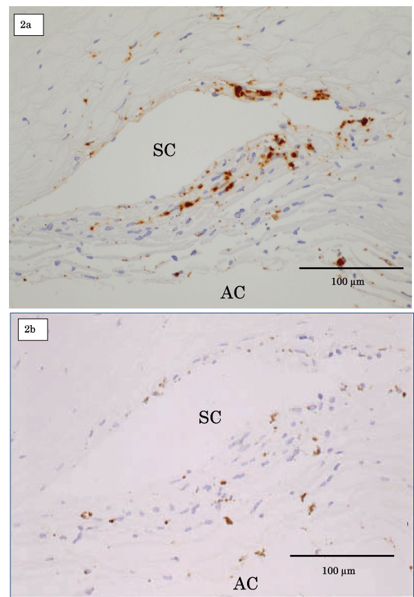
| Figure 2: Light microscopy of immunohistochemical staining, CD68 (2a) and control staining (2b) in Patient 5 (right eye). TLE was performed in the area of peripheral anterior synechia. CD68-positive cells were found around the Schlemm’s canal (SC), which was normally open. Although melanin-containing cells were seen, CD68-positive cells were negative in control staining. |