
|
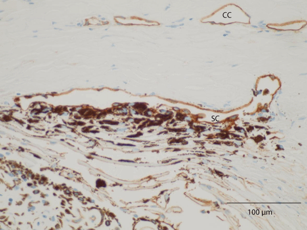
| CC: Collector Channels |
| Figure 4: Light microscopic photograph of a specimen subjected to thrombomodulin immunohistochemical staining, which can be used to detect Schlemm’s canal (SC). Trabecular cells in the uveal and corneoscleral meshwork were heavily pigmented. The most conspicuous finding was the existence of strongly pigmented trabecular cells in the cribriform meshwork. Note the absence of trabecular lamellae fusion and the normal size of the SC, which was positively stained with thrombomodulin |