|
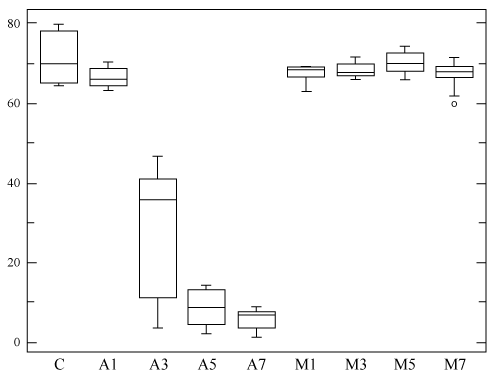
| Figure 3: Box plot showing the comparative effect of SA 1, 3, 5, 7 mM (A1, A3, A5, A7 respectively) and MEL 1, 3, 5, and 7 μM on Y79 cells in culture, as analyzed with the “Automated” (MuseTM, cell counter/analyzer - Merk Millipore)) method. C = Control, untreated samples. The “x” axis reports the increasing concentrations of both SA (A1 to 7) and MEL (M1 to 7). The “y” axis reports the percentage of live cells. SA 3, 5, and 7 mM cause extensive and statistically significant cell death in culture (see text) |