|
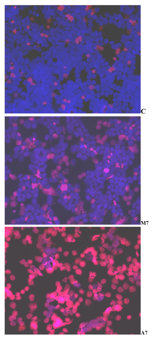
| Figure 4: Morphology of Y79 cells treated with MEL (M7), and SA (A7) and stained with Hoechst/PI. Cells were treated for 1 hour, harvested, washed, stained with Hoechst/PI, and examined by fluorescence microscopy, as described under “Material and Methods” (Original magnification 200 x) Blue nuclei (Hoechst 33342) indicate live cells, while red nuclei (Propidium Iodide) indicate dead cells. C = control (untreated) cells. 7 mM SA kills more than 90% Y79 cells in culture, as denoted by the red nuclei. Cell killing with MEL (M7) is not significantly different from the control (C). |