|
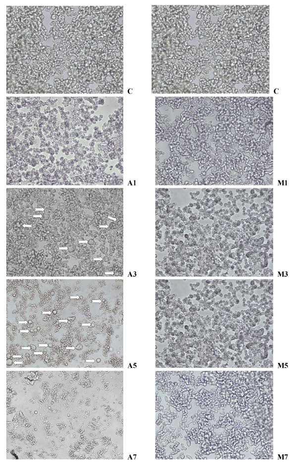
| Figure 5: Microphotographs of y79 retinoblastoma cells incubated with sodium ascorbate (SA) m 1, 3, 5, and 7 mM (A1, A3, A5, A7), and Melphalan (MEL) 1, 3, 5, and 7μM (M1, M3, M5, and M7). C = Control (untreated). Original magnification … 200 x). Cell treated with increasing doses of SA, show a progressive decrease in the total number of cells, especially evident in A7, and an increasing number of swollen cells (white arrows in A3, and A5) indicative of the phenomenon of “oncosis”. |