|
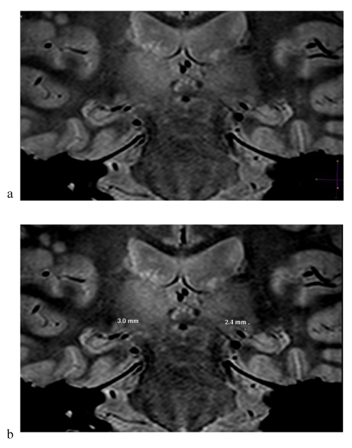
| Figure 1: LGN coronal slices in a patient with hypertensive glaucoma (M- 1942). a) Coronal 2 mm proton density weighted lateral geniculate nucleus image. b) Coronal proton density weighted LGN with oblique lines showing orientation used for height measurement. |