|
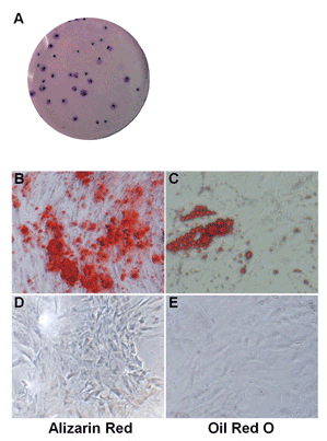
| Figure 2: Characterization of hASCs. A) hASCs were seeded at low density and incubated in CCM for 14 days to allow CFUs formation. The colonies were fixed and stained with crystal violet. B-C) representative images showing osteogenic (Alizarin Red staining) and adipogenic (Oil Red O staining) differentiation of hASCs, respectively. D-E) representative images showing hASCs cultured in CCM for 3 weeks as controls and stained with Alizarin Red and Oil Red, respectively. Original magnification 4x for B and D and 20x for C and E. |